Figure 6.
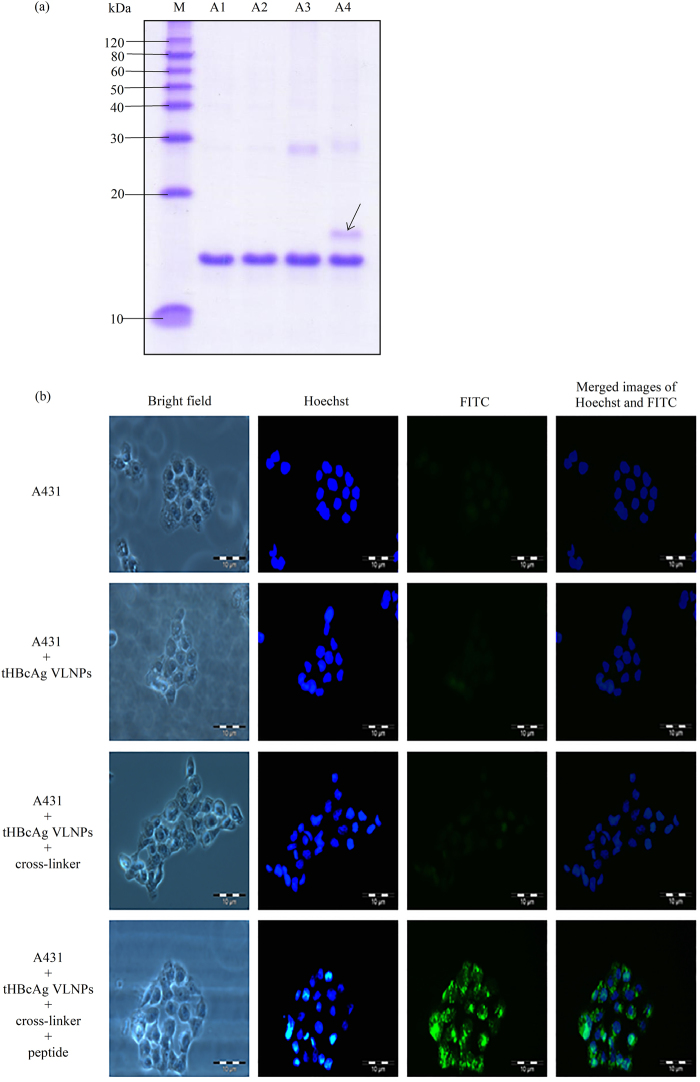
Delivery of tHBcAg VLNPs into A431 cells with peptide NRPDSAQFWLHHGGGSLLGRMKGA containing the nanoglue. (a) SDS-PAGE of tHBcAg conjugated to peptide NRPDSAQFWLHHGGGSLLGR-MKGA. Lanes M: molecular mass markers (kDa), A1: tHBcAg, A2: tHBcAg plus peptide NRPDSAQFWLH-HGGGSLLGRMKGA without cross-linker, A3: tHBcAg plus cross-linker without peptide, A4: tHBcAg plus cross-linker and peptide NRPDSAQFWLHHGGGSLLGRMKGA. The arrow shows the tHBcAg monomer conjugated to the peptide. The full length gel is shown in Supplementary Figure S1. (b) Delivery of tHBcAg VLNPs conjugated to peptide NRPDSAQFWLHHGGGSLLGRMKGA into A431 cells. tHBcAg was detected with the mouse anti-HBcAg monoclonal antibody, followed by the FITC-conjugated goat anti-mouse antibody. A431 cells incubated with tHBcAg VLNPs that had been conjugated to the peptide showed strong green fluorescent dots. The samples added to the cells are labelled on the left of figure.

