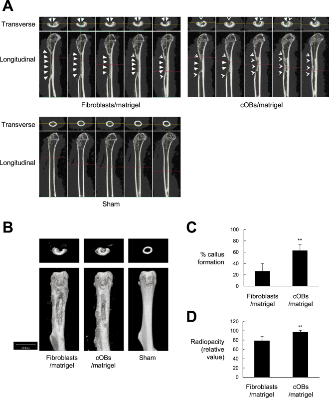
Figure 4.
cOBs facilitated bone repair in vivo. HDFs were cultured in osteogenic medium supplemented with ALK5 i II and VitD3 for 13 days (cOBs). The cOBs and the HDFs cultured in complete medium as control were inoculated into artificial segmental bone defect lesion in femoral diaphysis in NOG mice. Bone defect was not created in the sham-operated mice. Twenty-one days later, mice were sacrificed and μCT imaging of the femur was performed. (A and B), Longitudinal and transverse serial 100 μm slice images (A) and 3D-constructed μCT images (B) of the femurs of a representative mouse are shown. White triangles indicate bone defect lesions, while arrow heads indicate regenerated bone tissue. (C and D), The means ± S.D. of the percentages of callus formation (C) and the relative radiopacity at the bone defect region (value for the sham operated group are set to 100%) (D) are plotted (n = 4). **P < 0.01.

