Figure 5.
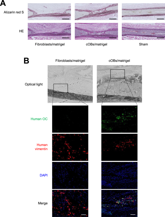
cOBs contributed to bone tissue regeneration in vivo. Transplantation experiment was performed as in Fig. 3. Twenty-one days after the cell inoculation, mice were sacrificed and the femur was excised. (A) Serial sections of the femur tissue were stained with Alizarin Red S (Upper) and H & E. (Lower). Scale bar = 1 mm. (B) Sections were stained with anti-human OC antibody followed by FITC-labeld secondary antibody, anti-human vimentin antibody followed by PE-labed secondary antibody, and DAPI. Optical light and fluorescence microscopic images are shown. Scale bar = 100 μm.

