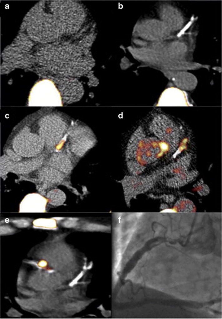
Figure 3.

Molecular imaging of coronary plaque osteogenesis via 18F-NaF PET imaging fused with coronary CT images. (A) Control patient with little coronary calcification or 18F-NaF uptake. (B) The panel demonstrates extensive coronary calcification without significant 18F-NaF uptake. (C and D) Focal NaF uptake in the LAD with flanking coronary arterial calcification. (E) Patient with a NSTEMI showing focal 18F-NaF tracer uptake in culprit lesion with (F) associated in-situ thrombus in the proximal right coronary artery. Reproduced by permission from reference (45).
