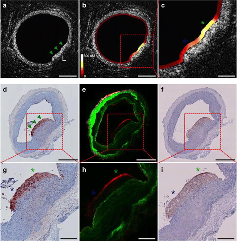
Figure 4.

High-resolution intravascular NIRF-OCT molecular-structural imaging of inflammatory protease activity in rabbit atherosclerosis. (a–c) Cross-sectional NIRF-OCT imaging of normal artery wall (a) and an atherosclerotic lesion (b,c), lipid-rich demonstrated by green and blue asterisks. (d–f) RAM-11 macrophage staining of plaque sections demonstrating high macrophage density, fluorescence microscopy with elevated plaque protease activity and positive cathepsin immunostain signal. (g–i) higher magnification of images d–f. Reproduced by permission from reference (70).
