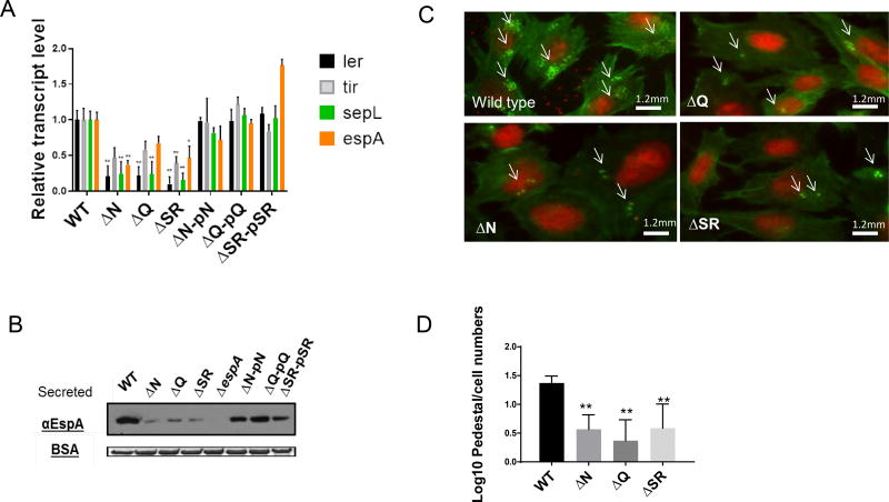
Figure 2. Lambdoid prophage genes control LEE expression in EHEC 86–24.
(A) qRT-PCR of LEE genes (ler, tir, sepL and espA) from RNA extracted from EHEC (WT, ΔN, ΔQΔΔSR, and complemented strains) grown under anaerobic conditions in DMEM low glucose. (n=6, error bars, standard deviation, P<0.01)
(B) Western blot for of the LEE-encoded and T3SS secreted protein EspA from secreted proteins of EHEC (WT, ΔN, ΔQΔΔSRΔespA and complemented strains) grown under anaerobic conditions in DMEM low glucose. Bovine serum albumin (BSA) was used as loading control.
(C) Fluorescent actin staining assay (FAS) depicting attaching and effacement (AE) lesions of EHEC (WT, ΔN, ΔQ and ΔΔSR) on infected HeLa cells. After 6 hours, cells were fixed, stained with FITC-phalloidin (actin in green) and propidium iodide (bacteria and HeLa DNA in red) and observed with fluorescence microscopy.
(D) Quantification of FAS, log transformation pedestal/cell numbers of WT EHEC, ΔN, ΔQ and ΔΔSR on Hela cells (n=150, error bars, standard deviation, P<0.01). Subjects with asterisks (**) indicates statistical significance at P<0.01 (Error bars, standard deviation, Student’s t-test). See also Figure S3.

