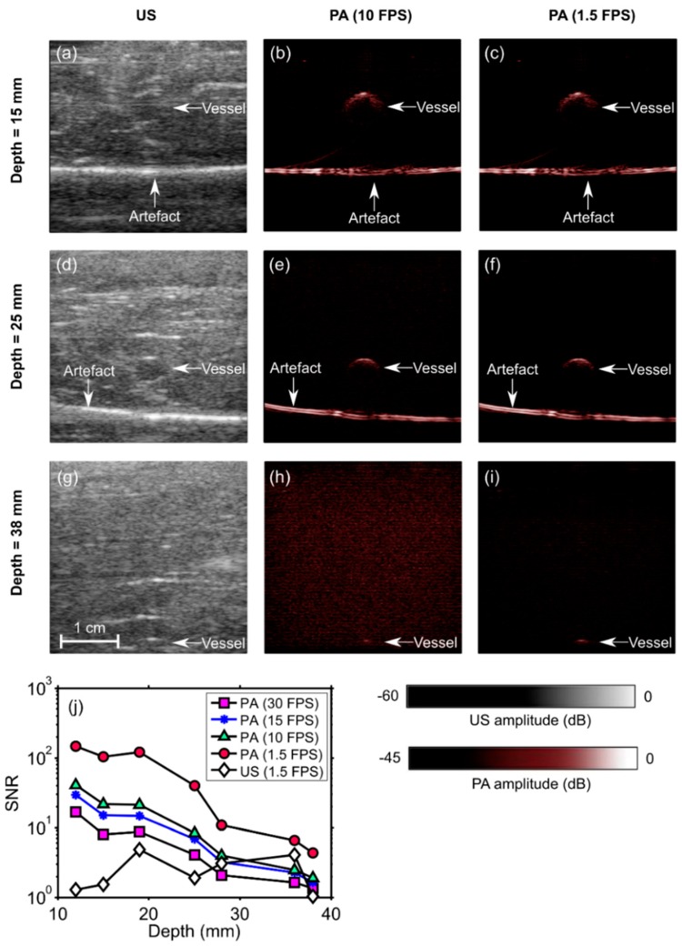
Figure 4.
Photoacoustic (PA) and ultrasound (US) images of a phantom comprising a vessel positioned in chicken breast tissue at different depths (a–i). The signal-to-noise ratio (SNR) of the PA images decreased with the vessel depth and with the imaging frame rate (j). At 1.5 frames per second (FPS), the SNR of the US images was lower than that of the PA images for all depths. During the insertions, these images were reconstructed and displayed in real-time on a logarithmic scale. Here, they are presented without the uppermost 5 mm, which contained the ultrasound gel. FPS: frames per second. Each point in the SNR plots was calculated from one spatial region.

