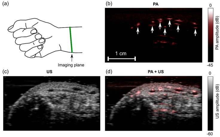
Figure 7.
Photoacoustic (PA) and ultrasound (US) images of a wrist of a human volunteer. (a) Schematic indicating the location of the imaging plane; (b) PA image; (c) US image; (d) PA + US image overlay. In the PA image (b), the prominent signals likely originated from blood vessels (arrows). Images were reconstructed and displayed in real-time on logarithmic scales. Here, they are presented without the uppermost 5 mm, which contained the ultrasound gel. Bulk tissue motion and localized pulsatile subsurface motion were apparent in the corresponding video (Supplementary Materials; Video S3).

