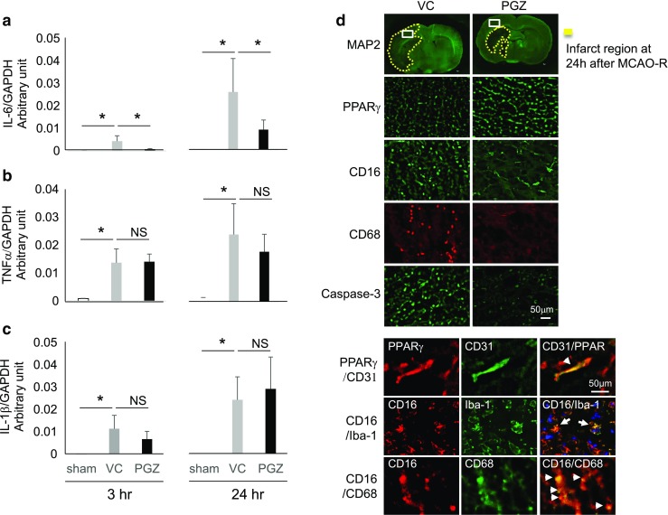
Fig. 2.

The mRNA level of pro-inflammatory cytokines and representative immunohistochemistry findings in PGZ- and VC-treated male rats. The mRNA level of the pro-inflammatory cytokines IL-6 (a), TNFα (b), and IL-1β (c) was assessed by quantitative real-time PCR assay and normalized by GAPDH. Data obtained 3 and 24 h after MCAO-R are shown. Data are the mean ± SD from eight rats per group. *p < 0.05 vs. VC by ANOVA followed by Scheffe’s test. The expression of PPARγ, CD16, CD68, and caspase-3 was examined in the peri-infarct region of PGZ-treated and VC rats 24 h after MCAO (d). PPARγ- and CD16-positive cells were co-localized with VEGF-, Iba-1- or CD68-positive cells
