Fig. 4.

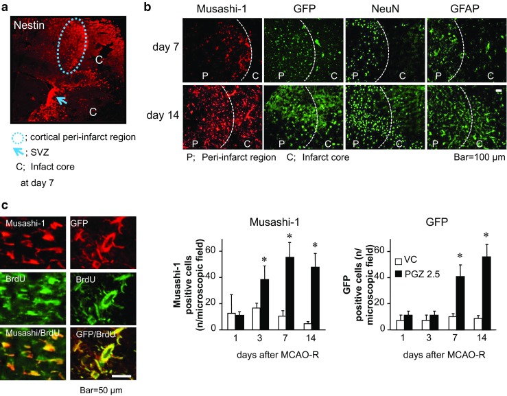
Stem cells migration from the SVZ into the cortical peri-infarct region promoted by PGZ in OVX/GFP+BM rats. Representative nestin-positive cells in the SVZ and the cortical peri-infarct region on day 7 after ischemia induction in PGZ-treated OVX/GFP+BM rats (a). Cells positive for Musashi-1, GFP, NeuN, and GFAP were detected in the infarct- and the peri-infarct area (b). Musashi-1- and GFP-positive cells were counted in 150 × 150-μm fields. The total cell number (n) in each area was 140–160 cells for DAPI. Each bar represents the mean ± SD from two areas in each of four rats. *p < 0.05 vs. VC by ANOVA followed by Scheffe’s test. Representative Musashi-1- and GFP-positive cells incorporated BrdU (c)
