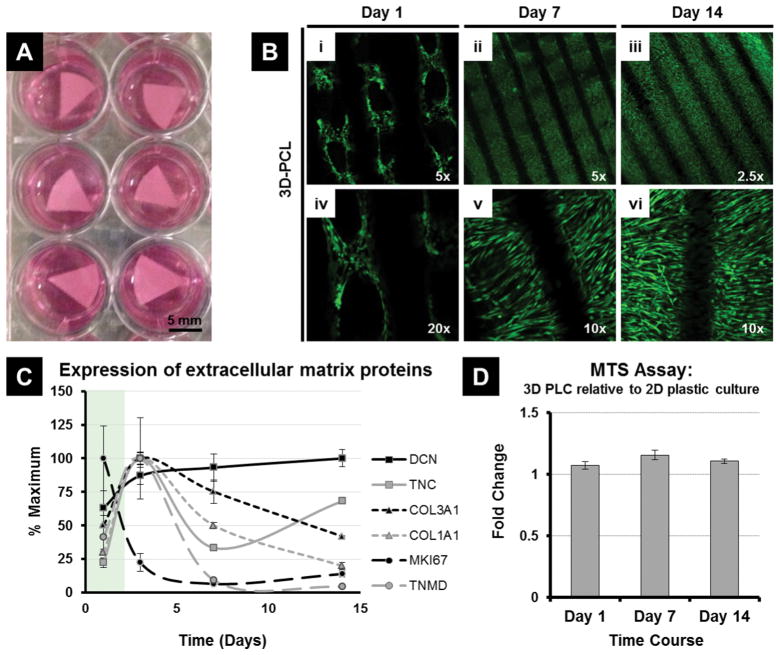
Figure 1.
Culturing Adipose Derived Mesenchymal Stem Cells (MSC) on 3D PCL Scaffolds. MSCs adhere, proliferate and are metabolically active on the scaffolds, with no indication of induced cytotoxicity. (A) PCL scaffolds seeded with MSCs and immersed in media containing 5% human platelet lysate. (B) Representative immunofluorescence microscopy images of MSCs cultured on 3D PCL scaffolds at day 1, day 7 and day 14 and stained using a cell viability assay (live cells shown in green, dead cells are stained red) to examine the survival, growth and morphology of the adherent cells. (C) Expression of key extracellular matrix proteins, as well as Decorin and the proliferation marker MKI67, in hPL cultured MSCs over 14 days. (D) Comparing the fold change in metabolic activity (via MTS assay) of MSCs cultured on 3D PCL scaffolds over 14 days relative to standard 2D plastic culture dishes. No significant difference (p>0.05) between treatment groups at each time point.

