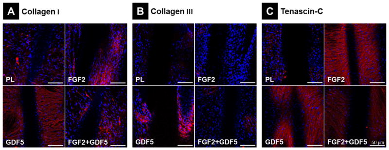
Figure 5.
Immunofluorescence Staining for ECM Cell Markers. Immunofluorescence staining (in red) of (A) Collagen 1, (B) Collagen III and (C) Tenascin-C to evaluate EMC protein accumulation in day 14 MSCs cultured on 3D PCL scaffolds according to 4 treatment groups. Blue = nuclear Dapi staining; scale bar = 50 μm.

