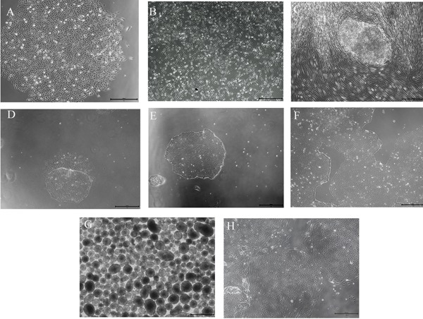
Figure 1.

Main types of cell culture (Scale bar = 500 μm). (A) Morphology of urine cells. (B) Urine cells on day 3 after electrotransfection. (C) iPSC colonies. (D) iPSC colonies after transition to feeder-free conditions with differentiated cells. (E) Purified colonies of iPSCs. (F) Stable growth and rapid proliferation of iPSC colonies are evident. (G) EBs are suspended. (H) Change in the morphology of EBs with adherence.
