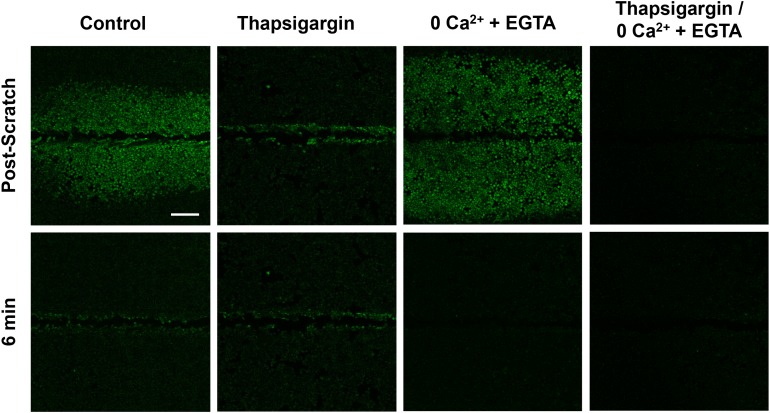
Figure 4. Signal propagation and persistence depend on distinct calcium stores.
Control groups showed scratch-induced increases in intracellular calcium at the wound edge that persist (measured up to 6 min), followed by propagating and transient calcium in neighboring cells (post-scratch vs. 6 min). Compared with control, intracellular calcium depleted (Thapsigargin) cells failed to propagate calcium signaling away from the wound edge. In contrast, depletion of extracellular calcium stores (0Ca2+ + EGTA) did result in signal propagation to neighboring cells but blocked persistent calcium at the wound edge. Depletion of both intracellular and extracellular calcium (Thapsigargin/0Ca2+ + EGTA) entirely blocked calcium signaling. Scale bar equals 200μm.

