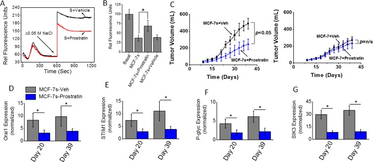
Figure 7.
(A) Fluo-3 Ca2+ measurement following Prostratin (8 μM) plus high salt treatment on MCF-7 cells. (B) Impact of high salt plus Prostratin (8 μM) treatment on intracellular Rhodamine-123 accumulation. (C) Tumorigenicity of high salt passaged breast cancer cells following oral administration of prostratin (100 μM). Temporal changes in the tumor volume following injection of 5 × 10⁵ MCF-7 and MCF-7s cells into Nu/J (n = 6) mice. (D–G) The mRNA expression of Orai (D), STIM1 (E), P-glycoprotein (F) and SIK3 (G). Data were represented as mean ± SEM, n = 6 per cohort, p < 0.05.

