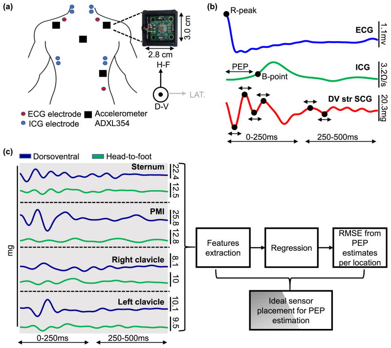
Fig. 2.
(a) The experimental setup for Part I of the experiment. Four ADXL354 Accelerometers are placed on the subject, one each at the mid-sternum, below the left and right clavicle, and at the point of maximal impulse. ICG and ECG signals are collected simultaneously. (b) Five beat ensemble averaged traces of ECG, ICG and mid-sternum dorsoventral SCG heartbeats. The ECG R-peak is used as a reference point for beat segmentation, the B-point of the ICG is used to detect aortic valve opening and the R-B interval is used as the ground truth PEP. Peak timing locations and width are extracted from the SCG signal as shown. (c) After extracting the features from the head-to-foot and dorsoventral axes of the SCG signals from all locations, a regression model is used to obtain PEP estimates from the features obtained from a single location, multiple combination of locations, one axis, and both axes. RMSE between the ground truth PEP and every estimate is calculated and the optimal location/ combination of location and axes is determined.

