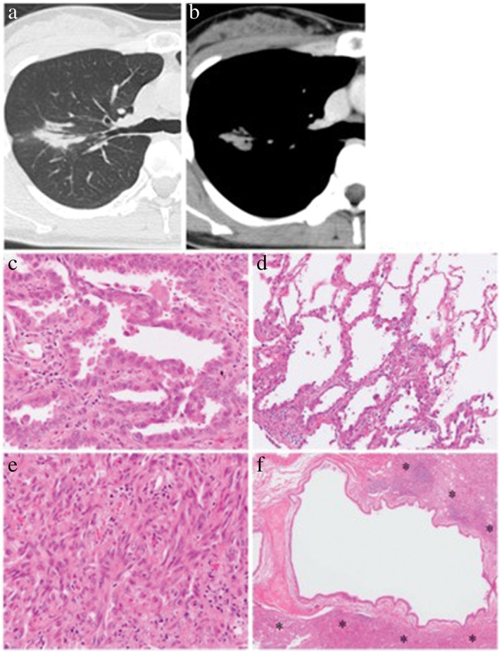
Figure 1.

Representative case of air bronchogram. (a,b) On thin‐section computed tomography scan, an air‐filled bronchus is surrounded by a lung tumor that only shows consolidation. (c–f) Histological examination of the resected tumor shows that this tumor is composed of (c) 60% papillary adenocarcinoma, (d) 30% lepidic adenocarcinoma, and (e) a 10% spindle cell component. (f) The intralesional bronchus remains intact, although it is surrounded by neoplastic tissue (asterisks). (c–f) Hematoxylin and eosin stain. Original magnification: (c,e) ×200, (d) ×100, (f) ×20.
