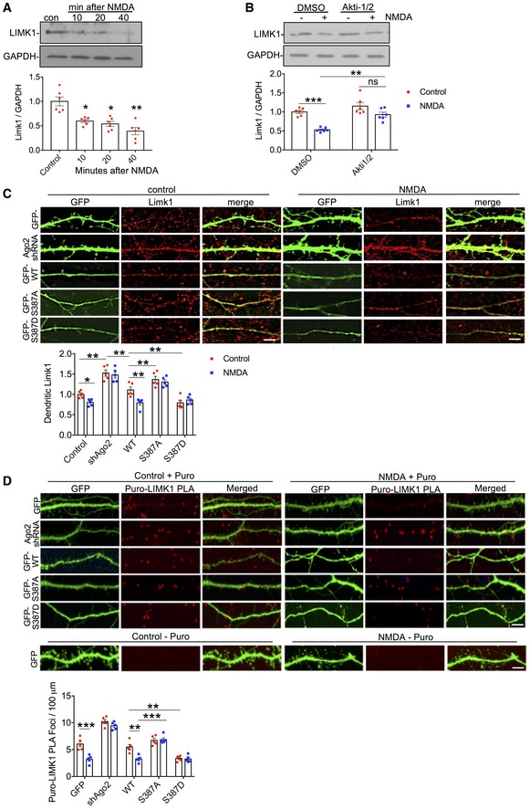
Figure 6. NMDAR stimulation regulates dendritic LIMK1 translation via Akt activation and Ago2 phosphorylation at S387.

- Endogenous LIMK1 protein levels are rapidly reduced in response to NMDAR stimulation. Cortical neuronal cultures were exposed to NMDA or vehicle for 3 min, and lysates were prepared 10, 20 or 40 min after NMDA washout and analysed by Western blotting. Graphs show quantification of LIMK1 expression normalised to vehicle control; n = 6. *P < 0.05; **P < 0.01; one‐way ANOVA, Bonferroni post hoc test. Mean ± SEM.
- NMDAR‐dependent decrease in LIMK1 is Akt‐dependent. Cortical neuronal cultures were treated with Akti‐1/2 20 min before NMDA or vehicle application, and lysates were prepared 40 min after NMDA washout and analysed by Western blotting. Graphs show quantification of LIMK1 expression normalised to vehicle control; n = 5. **P < 0.01; ***P < 0.001 one‐way ANOVA, Bonferroni post hoc test. Mean ± SEM.
- NMDAR‐dependent decrease in dendritic LIMK1 expression requires Ago2 phosphorylation at S387. Cortical neurons were transfected with molecular replacement constructs expressing Ago2 shRNA plus shRNA‐resistant GFP‐Ago2 (WT, S387A or S387D), fixed 40 min after NMDA washout, permeabilised and stained with LIMK1 antibodies (red channel). GFP signal was maximised at acquisition so that dendrites could be effectively visualised. Graph shows LIMK1 staining intensity in dendrites normalised to vehicle control; n = 5 independent experiments (10 cells per condition). *P < 0.05; **P < 0.01, two‐way ANOVA, Bonferroni post hoc test. Scale bar = 10 μm. Mean ± SEM.
- De novo translation of endogenous LIMK1 is regulated in dendrites by NMDAR stimulation via Ago2 phosphorylation at S387. Cortical neuronal cultures were transfected with molecular replacement constructs expressing Ago2 shRNA plus shRNA‐resistant GFP‐Ago2 (WT, S387A or S387D). Neurons were incubated with 1 μM puromycin in the presence or absence of 50 μM NMDA for 3 min. Following NMDA washout, neurons were incubated with puromycin for 40 min, after which the cells were fixed and processed for Puro‐LIMK1 PLA (see Materials and Methods). Puro‐PLA was also performed on GFP‐transfected neurons that were not incubated with puromycin as a negative control (bottom row of images). n = 5 independent experiments (10–13 cells per condition). **P < 0.01; ***P < 0.001 two‐way ANOVA, Bonferroni post hoc test. Scale bar = 10 μm. Mean ± SEM.
