
Figure 1.
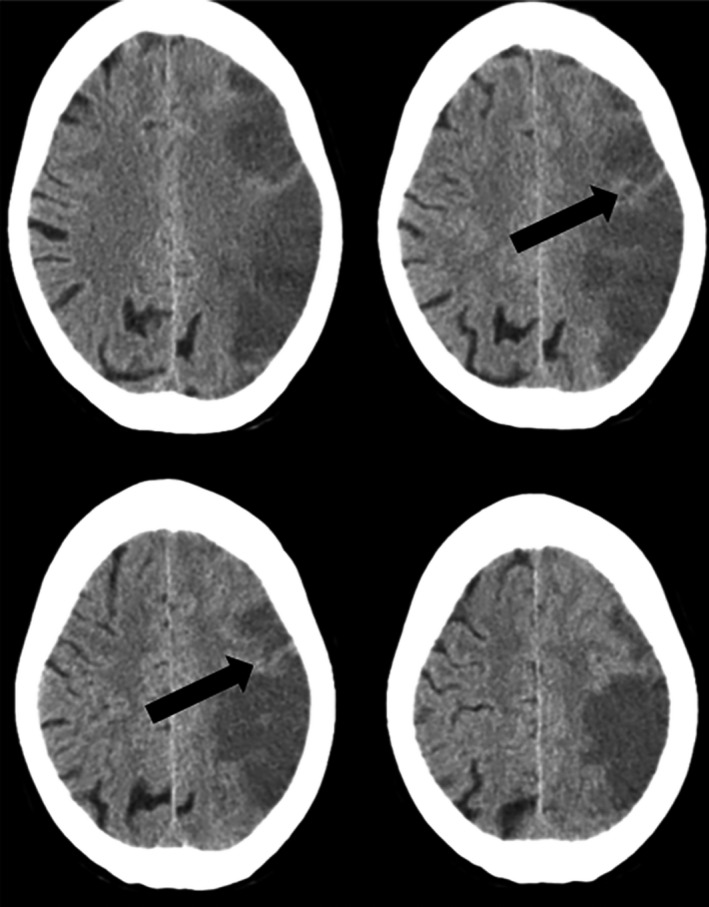
Islands of preserved cortex within the infarct. Brain CT scan showing an acute left middle cerebral artery ischemic stroke. Black arrows indicate cortical areas with normal attenuation coefficient (islands of preserved cortex) within the infarct.