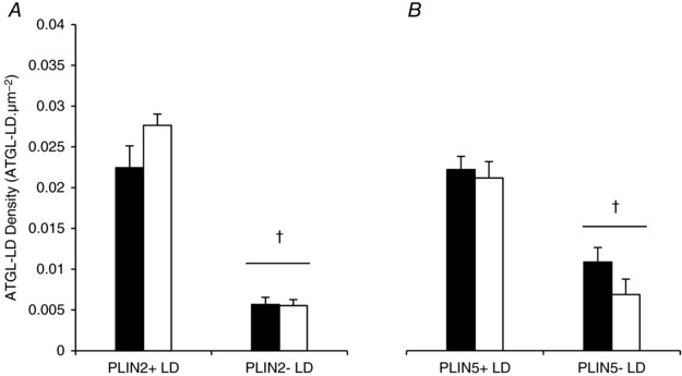
Figure 7. ATGL colocalisation with different pools of LDs does not change after exercise.

ATGL colocalisation with different LDs pools (PLIN+ or PLIN− LDs) was quantified from immunofluorescence microscopy images of ATGL, LDs and PLIN2 (A) or PLIN5 (B) before and after exercise. Data were collected as averages of 15 fibres per participant per time point. Values are presented as means ± SEM (n = 7 per group). †Main effect for PLIN association with LDs (P < 0.05 vs. PLIN− LDs).
