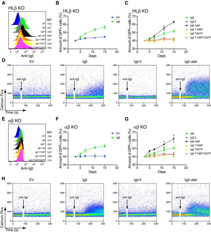
Figure 3. Signaling through the ITAM of BCR‐independent Igβ contributes to the fitness of Ramos cells.

-
AThe expression of Igβ on the surface of HLβ KO cells reconstituted with WT or different Igβ mutants was determined by flow cytometry.
-
B, CThe percentage of GFP‐positive transduced cells at different time points after the transduction of HLβ KO cells reconstituted with WT or different mutated form of Igβ is shown. The vectors express GFP as a transduction marker. The data represent the mean and standard error of a minimum of three independent experiments.
-
DCalcium responses of HLβ KO cells reconstituted with WT or different Igβ mutant constructs upon the stimulation of anti‐Igβ antibodies. The data are representative of three independent experiments.
-
EThe expression of Igβ on the surface of αβ KO cells reconstituted with WT and different Igβ mutant constructs was determined by flow cytometry.
-
F, GThe proportion of GFP‐positive αβ KO Ramos cells at different time points after their transduction with vectors encoding either WT or different mutated forms of Igβ. The vectors express GFP as a transduction marker. The data represent the mean and standard error of a minimum of three independent experiments.
-
HThe calcium responses of αβ KO cells reconstituted with WT or different Igβ mutant constructs stimulated by anti‐Igβ antibodies. The data are representative of three independent experiments. A minimum of two clones were used for both the HLβ KO and the αβ KO.
