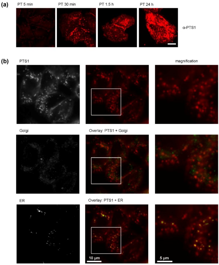
Figure 3.
(a) Time-dependent uptake of PT into CHO-K1 cells. CHO-K1 cells were incubated with PT (1 µg/mL). After the indicated time points, cells were fixed, permeabilized and blocked. PTS1 was detected with a specific monoclonal antibody. Pictures were taken with a Zeiss LSM-710 confocal microscope. Scale bar = 10 µm. (b) Co-staining of PTS1 with markers for Golgi or ER. CHO-K1 cells were incubated with PT (1 µg/mL) in the presence of ER-TrackerTM Blue White DPX (1 µM) for 3 h. Cells were fixed, permeabilized and blocked. PTS1 and GM-130 (Golgi marker) were detected by specific antibodies allowing the simultaneous detection of ER, Golgi and PTS1 in one sample. Left row of pictures shows the signal for the three single channels detected, middle row shows the PTS1 signal alone or in overlay with either Golgi or ER and the right row shows a magnification of the areas indicated by the white rectangle.

