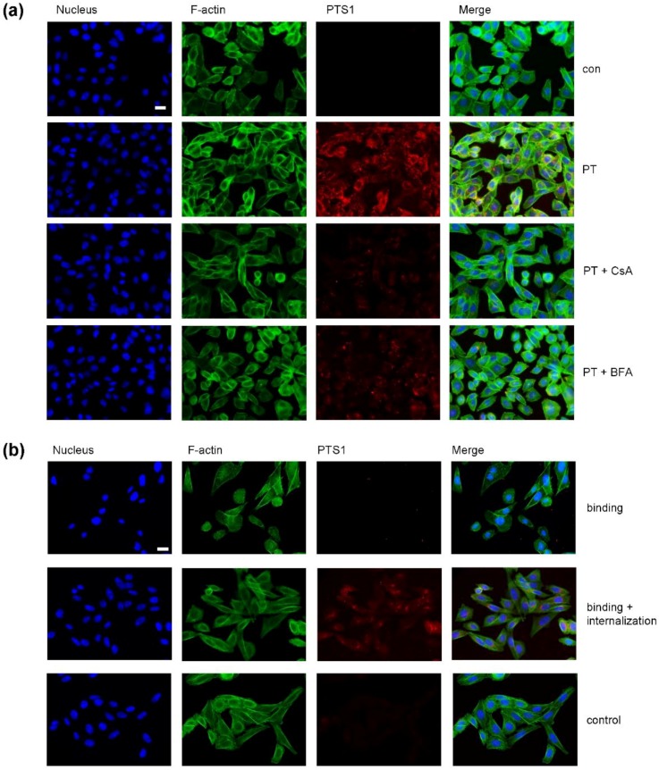
Figure 4.
(a) In the presence of CsA less PTS1 is detected in CHO-K1 cells. CHO-K1 cells were pre-incubated with CsA (20 µM) for 30 min. 20 µM BFA were used as control. Then, cells were challenged with 1 µg/mL PT in the presence or absence of the respective inhibitors and 3 h later cells were fixed, permeabilized and blocked. Then, cells were probed with an anti-PTS1 antibody, Hoechst and phalloidin-FITC for F-actin staining. Scale bar = 20 µm. (b) Anti-PTS1 antibody does not detect cell-bound PTS1. CHO-K1 cells were incubated on ice for 10 min and then challenged with 1 µg/mL PT for 30 min to enable only the binding to the cells. For control, cells were left untreated. Subsequently, cells were washed three times with PBS to remove unbound toxin. Then one portion of PT-treated cells was fixed immediately with PFA and another portion was further incubated at 37 °C for 2 h with subsequent PFA fixation. All samples were permeabilized, blocked and probed with a specific PTS1-antibody and a fluorescence-labelled secondary antibody. Nuclei were stained with Hoechst and F-actin with phalloidin-FITC. Scale bar = 20 µm.

