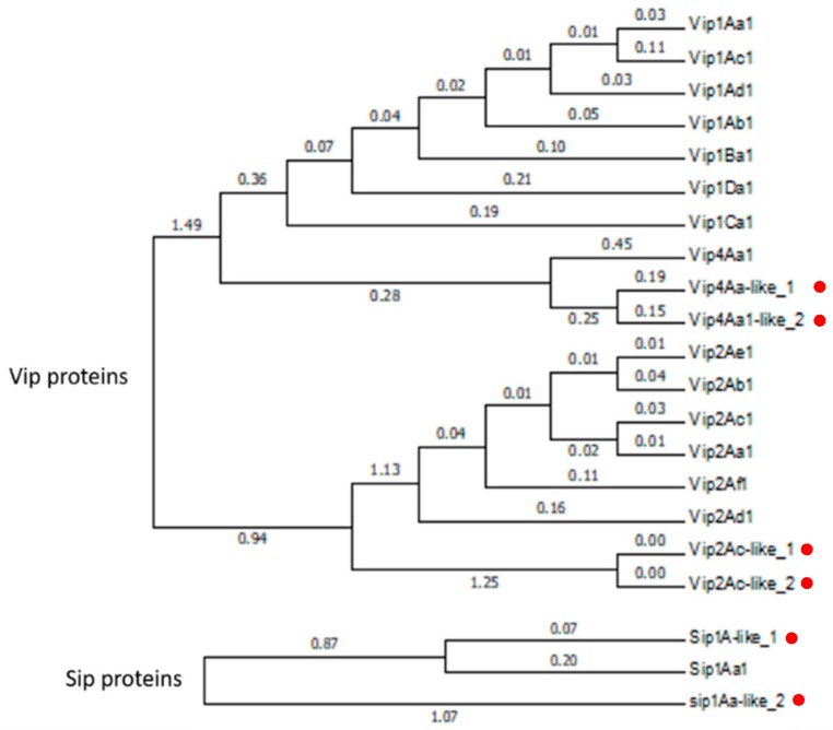
Figure 2.
Phylogenetic analysis of the Vip1/Vip2- and Sip1A-type proteins detected in the Bt isolates E-SE10.2 and O-V84.2. The red dots indicate the position of the new putative proteins in the phylogenetic tree. Branch lengths represent the number of substitutions per site of the multiple-sequence alignment as a measure of divergence (Mega v6 software).

