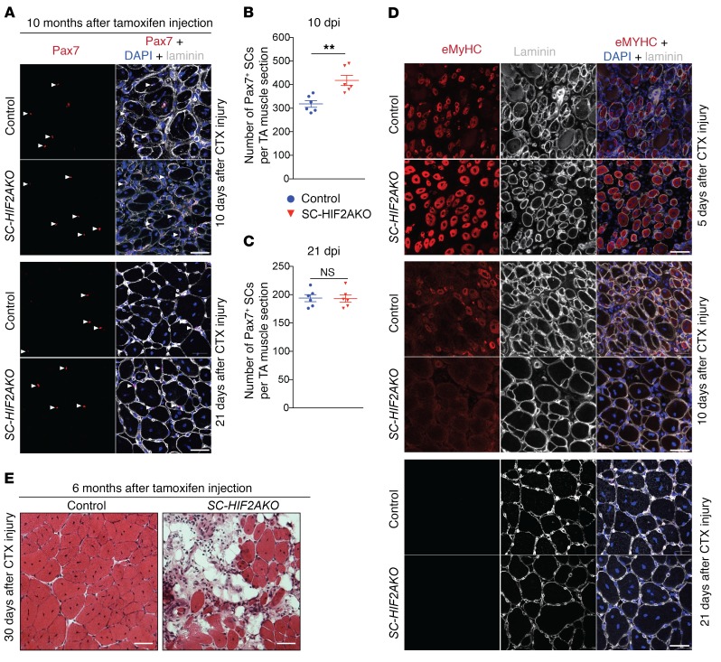
Figure 6. Genetic ablation of HIF2A transiently improves muscle regeneration but impairs long-term muscle regeneration potential.
(A) Representative images of TA muscles from SC-HIF2AKO mice and control littermates (n = 6 mice/group/time point). The muscles were CTX injured 10 days after tamoxifen-induced HIF2A ablation (10 dpr). Immunofluorescence of Pax7 after CTX injury (10 and 21 dpi) revealed an increase in Pax7+ SCs (arrowheads) in SC-HIF2AKO mice. Scale bars: 20 μm. (B and C) Number of Pax7+ SCs per TA muscle section at 10 dpi (B) and 21 dpi (C). (D) Representative images of TA muscles from SC-HIF2AKO mice and control littermates (n = 6 mice/group/time point). The muscles were CTX injured 10 days after tamoxifen-induced HIF2A ablation (10 dpr). Immunofluorescence of eMyHC and laminin B2 days after CTX injury (5, 10, and 21 dpi) revealed accelerated muscle regeneration in SC-HIF2AKO mice. Scale bars: 20 μm. (E) Representative images of TA muscles from SC-HIF2AKO mice and control littermates (n = 3 mice/group). The muscles were CTX injured 6 months after tamoxifen-induced HIF2A ablation. H&E staining of TA muscles 30 days after CTX injury revealed impaired muscle regeneration in SC-HIF2AKO mice. Scale bars: 20 μm. **P < 0.01, by 2-sided Student’s t test. Data represent the mean ± SEM.

