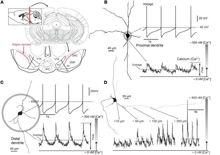
Figure 1. Combined patch clamp and Fura-2 Ca2+ imaging from SNc DA neurons revealed large oscillations in cytosolic [Ca2+] during pacemaking.
(A) Schematic coronal section of the midbrain, positioned 3.5 mm posterior to bregma. At the bottom, magnified ventral part of the midbrain showing sampled region of SNc (highlighted in red). (B) Whole-cell recording from a SNc DA neuron shown at left as a representative reconstruction of a Fura-2 –filled cell. At the bottom, 2PLSM measurement of Fura-2 fluorescence at a proximal dendritic location is shown (~15 μm from the soma). (C) Somatic recording during imaging at a distal dendritic location (~100 μm from the soma from the projection image of a SNc DA neuron). Note the increase in Ca2+ transient at the distal imaging site. (D) Ca2+ transients increased along the dendrite of shown SNc DA neuron ranging from 15 to 200 μm from the soma.

