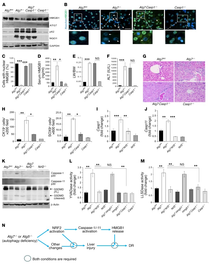
Figure 8. The inflammasome is involved in HMGB1 release in autophagy-deficient liver.
(A) Liver lysates from 9-week-old mice were analyzed by immunoblotting. (B) Liver sections were stained for HMGB1. Arrows indicate hepatocytes without nuclear HMGB1. Scale bars: 10 μm. (C) Cells with nuclear HMGB1 were quantified (n = 3 mice/group). (D) Serum levels of HMGB1 were measured. (E and F) LW/BW ratios (E) and serum ALT levels (F) were measured for 9-week-old mice. (G) Liver sections from 9-week-old mice were H&E stained. Original magnification, ×200. (H) CK19+ and SOX9+ cells were quantified. See also the images in Supplemental Figure 19A. (I and J) Hepatic mRNA levels of Casp1 (I) and Casp11 (J). Primer set 1 located in exon 6 was used for Casp11 amplification. (K–M) Liver lysates from 9-week-old mice were analyzed by immunoblotting or caspase activity assay (L and M). FL, full length. (N) Model of the role of NRF2, inflammasomes, and HMGB1 in liver injury, DR, and tumor development. NRF2 activation is required and sufficient for caspase-1 and caspase-11 activation and HMGB1 release in autophagy-deficient livers and is required, but may not be a sufficient condition, for autophagic liver injury or DR. Data represent the mean ± SEM. n = 3–7 mice/group. *P < 0.05, **P < 0.01, and ***P < 0.001, by 1-way ANOVA.

