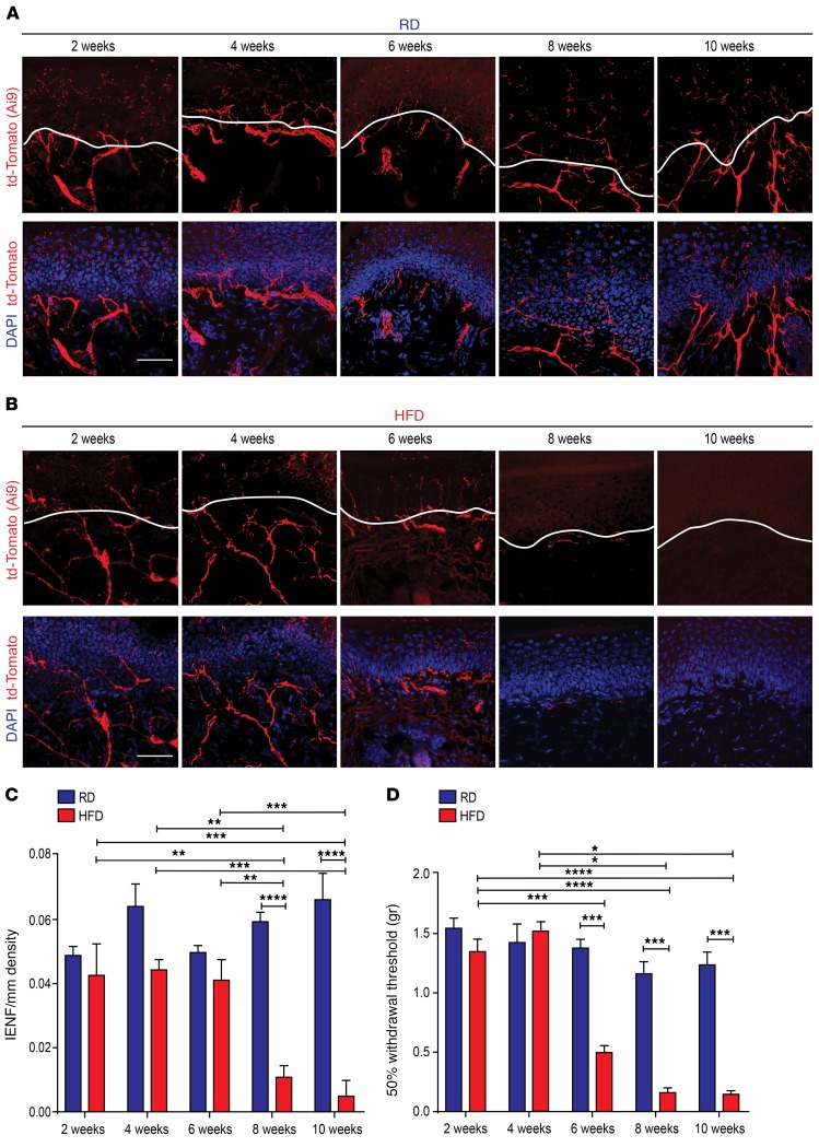
Figure 1. Onset of small-fiber degeneration and mechanical allodynia in mice fed a HFD.
(A) Confocal analysis of skin sections from Nav1.8-Cre;Ai9 mice fed a RD (blue) showed normal innervation. Nav1.8-positive fibers genetically labeled with td-Tomato are shown in red. Sections were stained with the nuclear marker DAPI (blue). Scale bar: 50 μm. (B) Skin sections from diabetic Nav1.8-Cre;Ai9 mice (HFD, red) had decreased innervation commencing 8 weeks after the start of the diet. Scale bar: 50 μm. (C) This effect was quantified using IENF density, and the epidermal-dermal junction is outlined in white in A and B. **P < 0.01, ***P < 0.001, and ****P < 0.0001 (n = 6 for all groups, with 3 noncontiguous sections analyzed per sample). (D) von Frey testing revealed the onset of mechanical allodynia in diabetic Nav1.8-Cre;Ai9 mice after 6 weeks on a HFD but not in RD-fed mice. *P < 0.05, ***P < 0.001, and ****P < 0.0001 (n = 7 mice/group). P values were calculated by 2-way ANOVA with Bonferroni’s multiple comparisons test. Values are expressed as the mean ± SEM.

