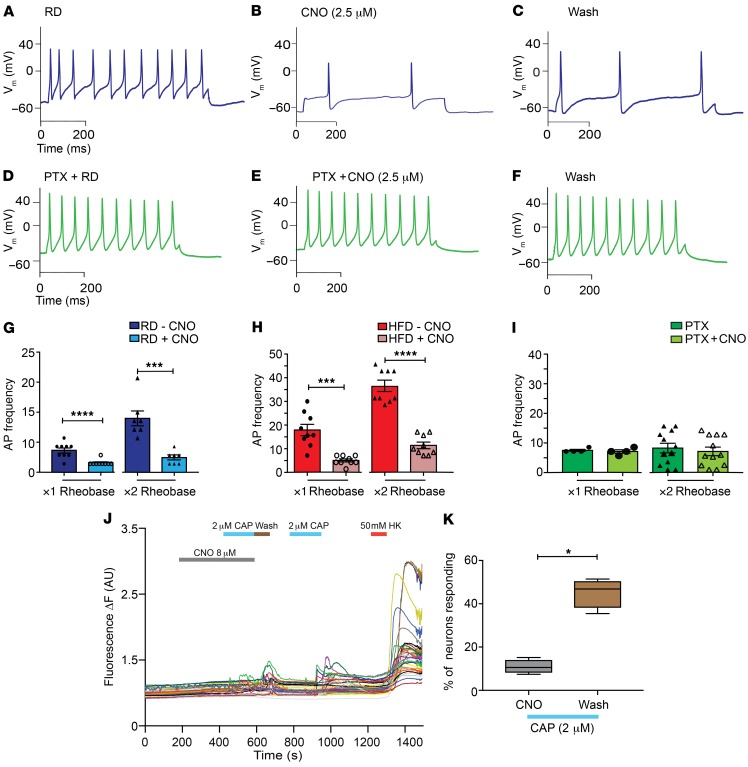
Figure 8. Chemogenetic inhibition of Nav1.8-positive DRG neurons expressing the inhibitory DREADD receptor PDi is G-protein mediated.
(A) Current-clamp recordings from inhibitory PDi–expressing Nav1.8-positive neurons in primary cultures isolated from Nav1.8-Cre;Ai9;RC::PDi mice fed a RD (blue). (B) Application of CNO (2.5 μM) reduced the AP frequency, and (C) washing out the CNO partially restored the firing rate. (D–F) Overnight incubation of RD DRG cultures with pertussis toxin (PTX, green) abolished the inhibitory effect of CNO. (G) In RD Nav1.8-positive DRG neurons expressing DREADD receptors, a significant decrease in AP frequency after application of CNO at both the ×1 and ×2 rheobase was observed. ***P < 0.001 and ****P < 0.0001 (n = 7 and 9, respectively). (H) The same mice fed a HFD also showed a decrease in AP frequency after application of CNO.***P < 0.001 and ****P < 0.0001 (n = 9 for both groups). (I) Overnight incubation of DRG cultures with pertussis toxin abolished the inhibitory effects of CNO. There was no difference in AP frequency after preincubation with PTX and application of CNO at either the ×1 or ×2 rheobase (n = 4 and 12, respectively). (J) [Ca2+]i responses in DRG explants from Nav1.8-Cre;RC::PDi GCaMP6 mice showed that [Ca2+]i responses after addition of capsaicin (2 μM) were inhibited during incubation with CNO (8 μM for 5 min). After washing, Nav1.8-positive DRG neurons showed restored [Ca2+]i transients to capsaicin (2 μM) and HK (10 mM) (n = 120 neurons; 10 explants). (K) The responses to lower concentrations of capsaicin were quantified as the responses to capsaicin as a percentage of the total number of HK-responsive neurons. *P < 0.05. Values are expressed as the mean ± SEM. P values were calculated using a Mann-Whitney U test.

