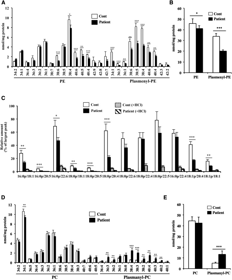
Fig. 4.
Quantification of ethanolamine and choline glycerophospholipids in the skin fibroblasts. Total lipids were extracted from the skin fibroblasts using the Bligh-Dyer method in the presence of internal standard, and the amount of each phospholipid was determined using LC-MS/MS. PE and plasmenyl-PE species (A) and total amount of ethanolamine phospholipids (B) scanned by monitoring the precursor ion of m/z 196 in negative mode. C: Plasmenyl-PE scanned by monitoring the precursor ions of m/z 364, 392, and 390 (fragment ions derived from plasmenyl-PE with 16:0, 18:0, and 18:1 at the sn-1 position, respectively) in positive mode. To eliminate plasmenyl-PE, samples were treated with HCl. PC and plasmanyl-PC species (D) and total amount of choline phospholipids (E) scanned by monitoring the precursor ion of m/z 184 in positive mode. Values were normalized against the amount of protein in the cells. Data are expressed as the mean ± SD from quadruplet analyses in one experiment. Each experiment was repeated at least three times with similar results *P < 0.05, **P < 0.01, and ***P < 0.001 as compared with control (Cont) cells.

