Fig. 8.
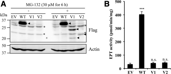
Expression of EPT1 variants in EPT1-KO HeLa cells. A: The EPT1-KO HeLa cells were transfected with empty vector (EV), vector containing Flag-tag fused WT EPT1 (WT), variant 1 (V1), and variant 2 (V2). After 24 h, cells were treated with 30 μM of MG-132 for 6 h (right panel). After cell lysates were separated by SDS-PAGE, Flag-tagged proteins were analyzed by immunoblotting using anti-Flag antibody. Actin was used as a protein loading control. Arrowheads and asterisks indicate Flag-specific and nonspecific signals, respectively. (B) EPT enzyme activity of the transfected cells treated with MG-132 was measured using radiolabeled CDP-ethanolamine as the substrate in the presence of diacylglycerol and Mn2+ for 10 min. ***P < 0.001 as compared with EV. n.s., not significant. Each experiment was repeated twice with similar results.

