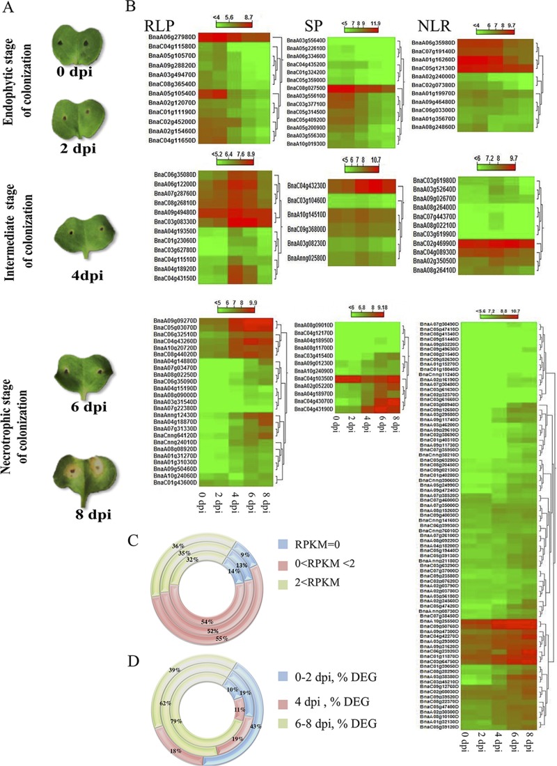
Fig 2. Symptoms and expression of candidate R genes after inoculation of susceptible Brassica napus cultivar Topas DH16516 with Leptosphaeria maculans isolate 00–100.
(A) Appearance of cotyledons 0, 2, 4, 6 and 8 days post-inoculation (dpi) with L. maculans. (B) Heat maps of differentially expressed genes (DEG) encoding receptor-like proteins (RLPs), secreted peptides (SPs) and nucleotide-binding leucine-rich repeat receptors (NLRs). The expression of each gene is based on regularized logarithmic transformation (rld) of the average of the best three biological replicates. Expression patterns are grouped into initial endophytic (0–2 dpi), intermediate (4 dpi) and late necrotrophic (6–8 dpi) stages of colonization [22]. (C, D) Classification of different gene families into different expression categories. Circles (from inside to outside) represent NLR, RLP and SP genes. (C) General expression patterns, reads per kilobase million (RPKM), did not differ between RLP, SP and NLR genes (S4 Table, χ2 = 2.51, P = 0.64). (D) Percentages of differentially expressed genes (% DEG) at the three stages of colonization are shown. Induced expression patterns differed between RLP, SP and NLR genes (S5 Table, χ2 = 22.13, P = 0.0002).

