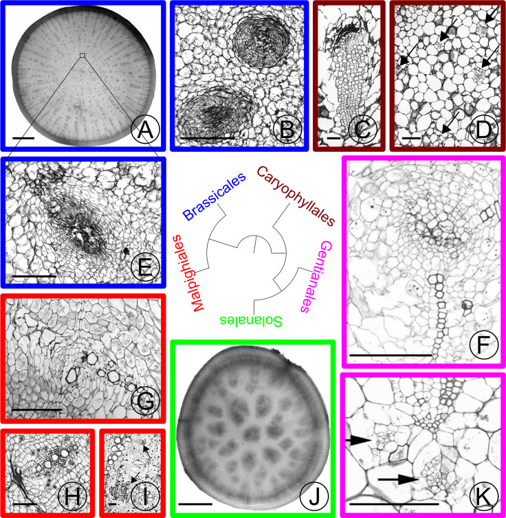
Fig 2. Stem and hypocotyl / root vascular bundles in parenchymatous tissues of various eudicot taxa.
Taxonomic orders are colored based on the label in the phylogeny. (A) Turnip (Brassica rapa rapa) cross section. Small dots in the center of the section are supplemental vascular bundles (SVBs). (B) Close-up of medullary SVB in the stem of kohlrabi (Brassica oleracea gongylodes cv. ‘Purple Vienna’). A small zone of xylem encircles crushed phloem in the amphivasal bundle arrangement. (C) Close-up of old medullary SVB from the trunk of Trichocereus chilensis (Cactaceae) showing a long tail of xylem adjacent to phloem that is capped with crushed secondary phloem (dark band). (D) Cross section of young stem just below the shoot apical meristem of Subpilocereus ottoni (Cactaceae) showing the distribution of five medullary bundles (arrows). (E) Close-up of SVB of turnip from boxed region in (A), showing the same arrangement of vascular tissues as kohlrabi. (F) Older medullary SVB from stem of Pachypodium namaquanum (Apocynaceae) with two zones of xylem and a zone of phloem interior to the xylem. (G) Old collateral medullary SVB from stem of Adenia keramanthus (Passifloraceae). (H) Old collateral SVB from root tuber of A. inermis. (I) Large collateral medullary SVBs from stem of A. metamorpha. (J) Cross section through tuberous root of Ipomoea batatas (Convolvulaceae). Dark regions in the center of the root are zones of proliferation with SVBs. (K) Medullary bundles from Caralluma burchardii (Apocynaceae) with two medullary phloem bundles (arrows) below protoxylem. Scale: (A), (J) 10mm; (B)-(I),(K): 250 m. (C) and (D) adapted from [4] with permission from the author; (F) and (K) adapted from [5] with permission from the author.

