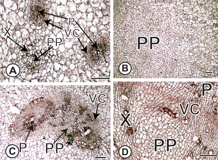
Fig 5. Anatomical evidence of proliferation of parenchyma (PP).
(A) PP within a VB in turnip in which proliferative cells are surrounded by scattered vessel elements. Two mature vascular bundles are labeled as well. (B) PP in center of kohlrabi stem. Note the profusion of smaller proliferative cells next to the larger, mature parenchyma cells. (C) PP has disrupted the patterning of a SVB. Note the fragments of phloem (darker staining) near the lighter-staining, small proliferative cells. (D) Intrusive proliferation of parenchyma near the main vascular cambium results in distorted cells and disrupted vessel elements. Note the sheared parenchyma cells next to the red-stained vessel elements that are no longer longitudinally oriented. From circled region in Fig 3F. Scale bars: 100μm. X = xylem; P = phloem; PP = proliferation of parenchyma; VC = vascular cambium.

