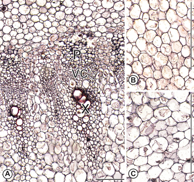
Fig 6. Anatomy of flowering kale and pak choi.
(A) Flowering kale vascular cambium and associated secondary xylem and phloem in flowering kale stem corresponding to the region with the small circle in Fig 3B. No intrusive growth is visible and cells are nearly isodiametric in contrast to the sheared cells near the turnip vascular cambium (Fig 5D). (B) and (C) Parenchyma cells in the vicinity of the small rectangles of Fig 3B and 3H of flowering kale and pak choi, respectively. Scale: A: 100μm; B, C: 400μm. X = xylem; P = phloem; VC = vascular cambium.

