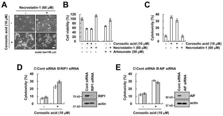
Figure 2.
Corosolic acid-induced cell death is independent of necroptosis. (A–C) Caki cells were treated with 10 µM corosolic acid or 50 µM artesunate (positive control) in the presence or absence of 60 µM necrostatin-1. We detected the cell morphology using interference light microscopy (A); XTT assay was used to detect the cell viability (B); LDH release assay was used to detect the cell cytotoxicity (C); (D,E) Caki cells were transiently transfected with siRNA against control, RIP1, and AIF. After 24 h, cells were treated with 10 µM corosolic acid for 24 h. LDH release was used to detect the cell cytotoxicity, and western blotting was used to detect the protein levels of RIP1, AIF, and/or actin. The values in the graphs (B,C,D,E) represent the mean ± SD of three independent samples.

