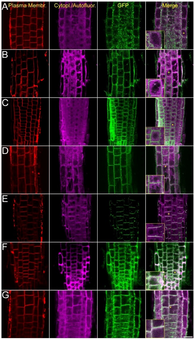
Figure 2.
Intracellular localization of the AtCRK-eGFP fusion proteins. AtCRK protein localization in root cells of transgenic Arabidopsis plants expressing the pCaMV35S::CRK-eGFP gene constructs. FM4-64 dye labeling indicates plasma membranes (red, first column). Violet laser induced (excitation: 405 nm) cytoplasmic autofluorescence is shown in the second column (pseudocolored in magenta, detected between 425–475 nm). GFP-conjugated CRK protein images (green, CRK1 to CRK8) are merged with autofluorescence images at the last column where vacuoles appear as dark intracellular areas due to lack of autofluorescence and absence of CRKs. Yellow asterisks indicate regions from which 2× magnified closeup images (insets) are prepared. (A) AtCRK1-GFP; (B) AtCRK2-GFP; (C) AtCRK3-GFP; (D) AtCRK4-GFP; (E) AtCRK5-GFP; (F) AtCRK7-GFP; (G) AtCRK8-GFP. Bar = 20 µm.

