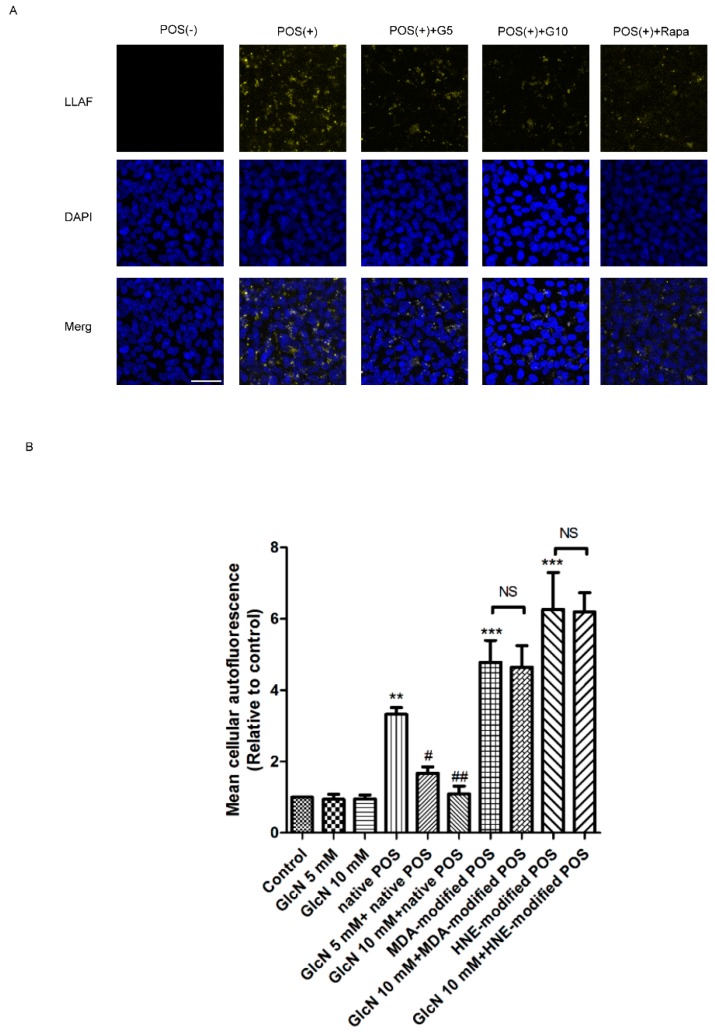
Figure 4.
Effects of glucosamine (GlcN) on photoreceptor outer segment (POS)-derived lipofuscin-like autofluorescence (LLAF) in ARPE-19 cells. (A) Effect of GlcN on native POS-derived LLAF in ARPE-19 cells. Cells were examined by confocal microscopy following seven days of treatment with native POS, native POS with GlcN (5 or 10 mM), or native POS with rapamycin (Rapa; 300 nM), and were compared with untreated cells. Magnification, ×200. Scale bar: 50 μM. (B) Effect of GlcN on either native or modified POS-derived LLAF in ARPE-19 cells. Cells were treated with GlcN (5 or 10 mM), native POS, native POS + GlcN (5 or 10 mM), MDA-modified POS, HNE-modified POS, MDA-modified POS + GlcN (10 mM), or HNE-modified POS + GlcN (10 mM), and were compared with untreated cells. The mean fluorescence intensity (MFI) was quantified by flow cytometry following seven days of incubation. The results are represented as mean ± SEM. The differences of the MFI in ARPE-19 cells were compared between groups using ANOVA, and Tukey’s test was used for post hoc analysis; ns, not significant; ** p < 0.01 versus the control group; *** p < 0.001 versus the control group; # p < 0.05 versus the native POS group; ## p < 0.01 versus the native POS group.

