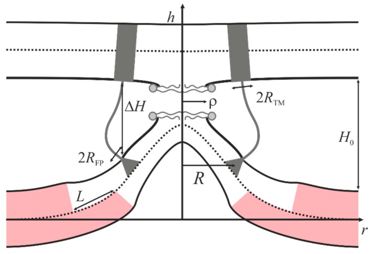
Figure 3.
Schematic representation of the model. Distance ΔH between fusion peptides and transmembrane domains of proteins in membranes is used as the reaction coordinate. Transmembrane domains (half-width of RTM) are schematically shown by gray rectangles, fusion peptides (half-width of RFP) are shown by gray triangles. H0 is the equilibrium distance between the membranes, ρ is the radius of the hydrophobic face formed in the area of maximal proximity of the membranes. Raft area is highlighted in pink. The viral membrane is shown on the top, and the cellular membrane is shown at the bottom.

