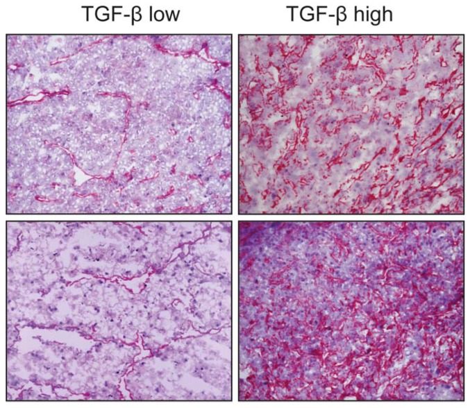
Figure 3.
Expression of transforming growth factor-β (TGF-β) in 4 HCC samples. Overall staining is evident, with a preferential sub-endothelial localization, possibly perisinusoidal, especially in tissues with lower expression. The cytokine delivery sites appear to be closely associated to parenchymal cells in HCC nodules. Representative images at 10×.

