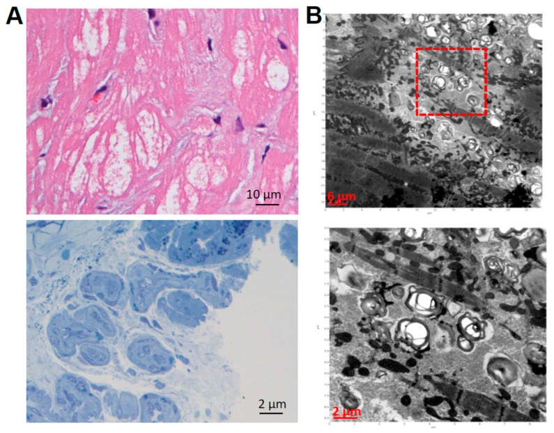
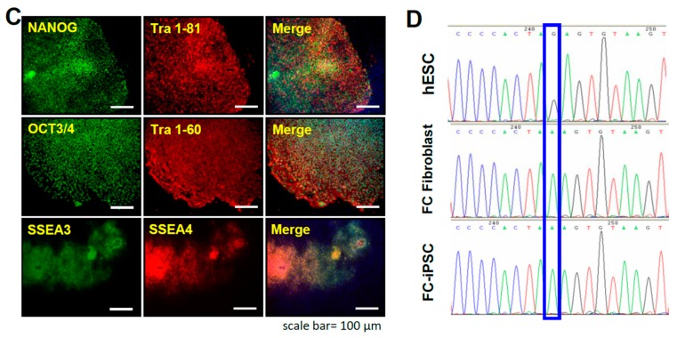
Figure 1.
Generation of FC-iPSCs. (A) Patients who carry intervening sequence (IVS) +919 G>A mutation and marked ventricular hypertrophy were assigned for myocardial biopsy. In the biopsied myocardial specimen, H & E staining showed the obvious hypertrophy and disorganization of cardiomyocytes containing large perinuclear sarcoplasmic vacuoles (upper). The toluidine blue staining verified the accumulation of glycosphingolipid in the myocardium (lower); (B) Transmission electron microscope (TEM) examination detected the formation of lamellar bodies (Zebra bodies) that represented glycolipid-containing lysosomes × 60,000 (lower). Red square indicated the abnormal lysosomes containing glycolipids. H & E staining, toluidine blue staining, and TEM examination (A,B) are representative results from a patient with IVS4+919 GLA G>A mutation and cardiomyopathy; (C) Immunofluorescence indicated the pluripotency markers Oct3/4, Nanog, SSEA3, SSEA4, TRA-1-60 and TRA-1-81 in FC-specific iPSCs (FC-iPSCs) clones. Scale bar, 100 μm; (D) Sequence analysis confirmed the existence of the specific IVS4+919 G>A mutation in patient-specific FC-iPSCs. Blue rectangle indicated the G>A mutation detected by sequence analysis. Results indicated the representative IVS4+919 G>A mutation in a patient-derived FC-iPSC clone.


