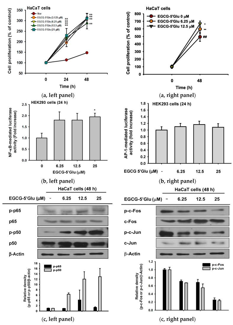
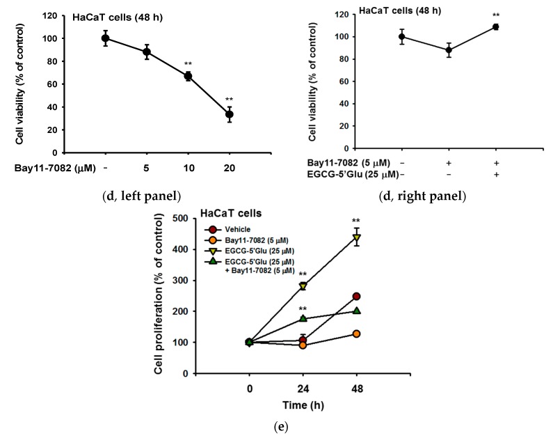
Figure 5.
Effect of EGCG-5′Glu on cell proliferation. (a) Proliferation of HaCaT cells treated with EGCG-5′Glu (0–25 μM) for 0–48 h was measured by MTT assay (left panel) and by Trypan blue dye exclusion assay. (b) HEK293 cells were transfected with NF-κB-Luc (left panel), AP-1-Luc (right panel), and β-gal plasmids and treated with EGCG-5′Glu (0-25 μM) for 24 h. (c) Levels of phospho- and total forms of p65 and p50 (left panel) and c-Jun and c-Fos (right panel) in whole cell lysates were determined by immunoblot analysis after treating HaCaT cells with EGCG-5′Glu (0–25 μM) for 48 h. (d, left panel) Viability of Bay11-7082-treated HaCaT cells was measured by MTT assay for 48 h. (d, right panel) Bay11-7082 (5 μM) was treated with or without EGCG-5′Glu (25 μM), and viability of HaCaT cells was measured by MTT assay. (e) With EGCG-5′Glu (25 μM) treatment, the effect of NF-κB on cell proliferation using Bay11-7082 (5 μM) was confirmed by MTT assay. * p < 0.05 and ** p < 0.01 versus a control group (normal group).


