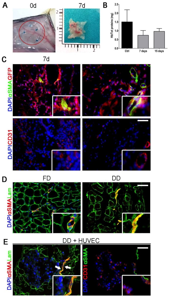
Figure 2.
Scaffold subcutaneous implantation and vessel after DET treatment. (A) Macroscopic appearance of the implanted DD at day 0 (red circle) and of DD explanted after 7 and 15 days of implantation; (B) Quantification of hemoglobin over total protein content in control (skin) and DD after 7 and 15 days of implantation (mean ± SD); (C) Immunofluorescence staining against αSMA and GFP performed in explanted DD after 7 and 15 days from implantation (upper panel) and CD31 (lower panel); nuclei were counterstained with DAPI; (D) Immunofluorescence staining against αSMA and Laminin performed in fresh diaphragm (FD) and DD; nuclei were counterstained with DAPI; (E) Immunofluorescence staining against αSMA and Laminin or CD31 performed in DD after 48 h of incubation with HUVECs; nuclei were counterstained with DAPI; Scale bar = 100 µm.

