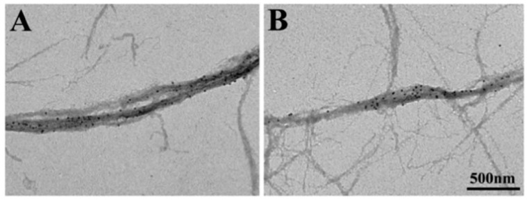
Figure 11.
Conventional electron micrographs showing part of actin and myosin filament mixture. Myosin heads were position-marked with antibody 1 (left) and antibody 2 (right). Note that thick myosin filaments are surrounded by thin actin filaments. From Sugi et al., [10].

