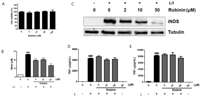
Figure 6.
Effect of robinin on inflammatory responses in mouse peritoneal macrophages. (A) Mouse peritoneal macrophages were cultured with robinin for 24 h. Cell viability was determined using the MTT assay. Data are represented as percentage of control cells (0 μg/mL robinin) (n = 4). (B,C) Cells were stimulated with LPS (L) and IFN-γ (I) in the presence of robinin for 24 h. The level of iNOS protein (B) was analyzed by Western blotting using tubulin as an internal control. One of three independent experiments is shown. The levels of nitrite (C) were measured using the Griess reaction. (D,E) The levels of TNF-α at 6 h (D) and 24 h (E) in the supernatant was measured by ELISA (n = 3). ### p <0.005 vs. control (−L); *** p < 0.005 vs. control (+L/I).

