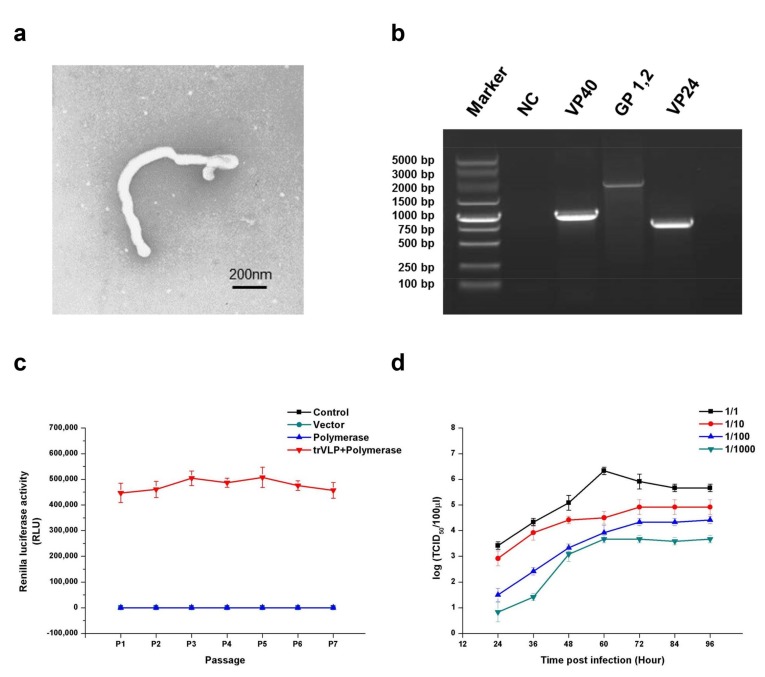
Figure 1.
Morphological, genetic, and stability identification; stability analysis; and proliferative ability determination of transcription- and replication-competent virus-like particles (trVLPs). (a) Electron microscopy of trVLPs. P1 trVLPs were concentrated using centrifugal filter devices (Millipore Corporation, Temecula, CA, USA) and then observed by electron microscopy. Scale is 200 nm. (b) Genetic identification of VP40, GP, and VP24. Reverse transcription polymerase chain reaction (RT-PCR) was performed to obtain the complete sequences of the three genes in the minigenome. The products of RT-PCR were identified by sequencing. NC: Cells with no trVLP infection. (c) Renilla luciferase activity from P1 to P7. To evaluate its hereditary stability, the trVLPs containing the recombinant virus genome were carried on to successive generations, and the Renilla luciferase activities from P1 to P7 were detected. In particular, the Renilla luciferase activities of the three groups (control, vector, and polymerase) were almost at the same level. The products of RT-PCR for the trVLP genome from P0 to P7 were identified by sequencing. The data of this sequencing experiment confirmed that the sequences of the trVLP genome were the same as those of wild-type EBOV (Zaire Mayinga Ebola virus) and the sequences remained unchanged from P0 to P7. (d) Proliferation curve of trVLPs in 293T cells. The virus proliferation curve in 293T cells was constructed by detecting the 50% tissue culture infective dose (TCID50) of the trVLPs from the cell culture supernatant. The TCID50 of trVLPs used for infection were 10−6.5/mL, 10−5.5/mL, 10−4.5/mL, and 10−3.5/mL from 1/1 to 1/1000, accordingly. The results in (c,d) are presented as the mean ± SD of three independent experiments.

