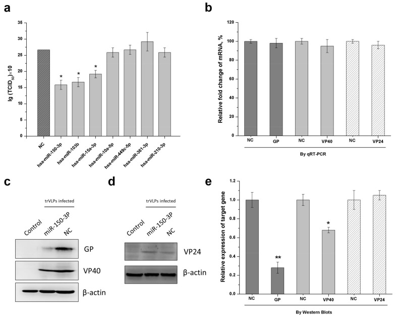
Figure 3.
miR-150-3p inhibits the reproduction of trVLPs by inhibiting GP and VP40 expression. 293T cells were transfected with support plasmids and a miRNA-expressing plasmid, and then infected with trVLPs. (a) Detection of the inhibitory effect from each miRNA is shown in Table 1. The best inhibitory effect against trVLP was achieved by miR-150-3p, as determined by the TCID50 of the cell culture supernatant 72 h post infection. NC: empty vector was used as a control. (b) At 72 h post infection, the total RNA was extracted, reverse-transcribed, and quantified by qPCR. Relative fold changes in mRNA were compared with infected, non-miR150-3p-expressing cells (NC). Charcoal grey represents the mRNA fold change of GP; light grey represents the mRNA fold change of VP40; white with slashes represents the mRNA fold change of VP24. The protein levels of GP, VP40 (c), and VP24 (d) were analyzed by Western blotting. β-actin was used as an internal reference. Control: cells with no infection; NC: cells with infection; miR-150-3p: infected cells with the expression of miR-150-3p. (e) The intensities of the respective images in (c,d) were quantified using ImageJ software. Charcoal grey represents the relative expression of GP; light grey represents the relative expression of VP40; white with slashes represents the relative expression of VP24. The results in (a,b,e) are presented as the mean ± SD of three independent experiments; * p < 0.05; ** p < 0.01.

