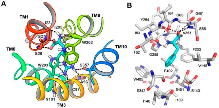
Figure 10.
(A) Periplasmic view of the substrate binding site of AdiC (PDB ID: 3L1L). Arginine (cyan) is bound to AdiC at the center of the transport path, recognized by amino acids from TM1, TM3, TM6, TM8, and TM10. Arginine and interacting residues of AdiC are shown in stick representation; (B) Predicted binding mode of phenylalanine in LAT1. LAT1 (gray) and phenylalanine (cyan) are shown in stick representation. (B) Figure adapted after securing permission from reference [44].

