Figure 1.
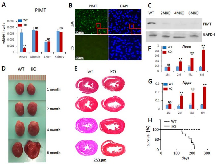
Cardiac-specific ablation of PIMT expression causes dilated cardiomyopathy. (A) Quantification of PIMT mRNA relative to 18S ribosomal RNA by RT-qPCR in PIMTfl/fl (WT) and csPIMT−/− (KO) mouse heart, muscle, liver and kidney; (B) Immunohistochemical localization of PIMT in 2-month-old PIMTfl/fl (WT) and csPIMT−/− (KO) mouse hearts. Nuclear localization of PIMT is evident in WT but not in KO hearts; compare DAPI stained images shown in right; (C) Western blot analysis for detecting PIMT protein level in PIMTfl/fl and csPIMT−/− mouse heart homogenates; (D) Representative photographs of heart of 1-, 2-, 4-, and 6-month-old csPIMT−/− mice and their PIMTfl/fl littermate controls. Six-month-old csPIMT−/− mouse hearts were flaccid and flabby; (E) Cross sections of hearts shown in Figure 1D were stained with H&E to reveal thinning of ventricular walls and dilation of chambers in csPIMT−/− mouse hearts (right panel); (F,G) Nppa and Nppb mRNA levels, respectively, in PIMTfl/fl and csPIMT−/− mouse hearts obtained at indicated ages. Each group was analyzed using 5 different mice (each mouse was assayed separately) and the values were expressed as the mean ± SD. * p < 0.05, ** p < 0.01, NS: not significant; (H) Survival curve showing lethality of mice with csPIMT−/− hearts. 36 mice for each group of PIMTfl/fl and csPIMT−/− were used for the generation of survival curve. Kaplan-Meier method was used to determine the survival rates and data were compared using log rank test. Each group was analyzed using 5 different mice and the values were expressed as the mean ± SD. * p < 0.05, ** p < 0.01, NS: not significant.

