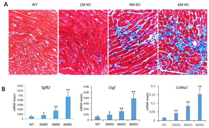
Figure 5.
Myocardial fibrosis in csPIMT−/− mouse hearts. (A) Images of Masson trichrome staining patterns of a representative PIMTfl/fl and csPIMT−/− hearts of 2, 4 and 6 months are shown (magnification 400×). Note the intensely stained (blue color) interstitial fibrous strands in 6-month-old csPIMT−/− hearts; (B) Quantification of mRNA levels for Tgfβ2, Ctgf and Col9a2 in csPIMTfl/fl and csPIMT−/− hearts of 2, 4 and 6 months of age. mRNA levels were quantified by RT-qPCR assays. Each group was analyzed using 5 different mice and the values were expressed as the mean ± SD. * p < 0.05, ** p < 0.01.

